Friday, October 26, 2012

Cervical Fusion Surgery

Cervical Fusion Surgery

Fusion of the Neck Vertebrae

By , About.com Guide
Updated September 20, 2011
About.com Health's Disease and Condition content is reviewed by the Medical Review Board
 
File for SSI DisabilitySocialSecurityLawFirms.com/FormHelpFind Out If You Qualify For Social Security Benefits. Free Evaluation!
Cervical Spinal Stenosiswww.SafeSpineSurgery.comTrust The Neuro-Ortho Specialists Best Of NYC Dr's Stern and Neubardt
A "cervical fusion" is the name for a surgical procedure that links together damaged segments of the vertebral column in the neck. This surgery is usually required when the cervical vertebrae -- and the discs between each vertebrae -- have become damaged as a result of an injury or chronic wear-and-tear. During surgery, the disc(s) between one or more vertebrae are removed, and bone growth is stimulated to link together adjacent vertebrae. Often, a metal device is used to stabilize the fusion until the bone growth is solid.

Cervical Fusion Surgery

A cervical fusion, also called an arthrodesis, permanently links together two (or more) adjacent vertebrae. Normally, there is a spinal disc between each vertebrae. The disc acts as a cushion, but also allows some movement between each vertebrae. Most often a cervical fusion is performed because the spinal disc is causing problems by pushing on a nerve (called a disc herniation). This nerve pressure can cause irritation leading to symptoms of pain, tingling, and numbness in the neck and arms. When a cervical fusion is performed, the disc, or fragments of the disc are removed. The bones of the vertebrae are then permanently linked together. This linkage occurs both with bone graft (the permanent solution) and often a metal plate, screw, or rod (the temporary solution). The metal simply holds the vertebrae in position while bone permanently fuses the segments together. Once the bone has linked the two vertebrae together, the fusion is considered solid, and resumption of normal activities is allowed.

Recovery From Cervical Fusion

Recovery from cervical fusion depends on a number of factors. As mentioned, the surgery is considered a success when the symptoms are improved, and the bone has healed across the fused vertebrae. This fusion process usually takes 2 to 3 months. The activity allowed during that time will depend on the strength of the fusion. In some patients with solid bone and strong metal fixation more activity may be allowed. On the other hand, individuals who have concerns about their ability to fuse the vertebrae, the recovery may be more cautious.

Complications From Surgery

The most common complication from surgery is basically that it fails to relieve the persistent neck pain. Fortunately, these complications are not common, but can occur. Studies have found success rates between 80 to 90% with cervical fusion surgical treatment for radiculopathy (nerve pain). The other potential major complication of a cervical fusion is a lack of adequate bone growth between the adjacent vertebrae. This is called an incomplete fusion, and may require additional surgery. Bone may not grow adequately for a number of reasons, and doing things like avoiding smoking and not taking medications or drugs that are known to interfere with bone growth. Other factors (such as a person's natural bone strength) are difficult to change. Other complications of cervical fusion can include nerve injury, difficulty swallowing, infection, and bleeding. Many patients are concerned about injury to the spinal cord. Of all of the complications listed, this is probably the least common. The risk of spinal cord injury is a small fraction of a percent.

Alternatives to Cervical Fusion

If a patient only has a small disc herniation, often just the disc fragment can be removed without requiring a fusion. But if a patient needs the full-blown surgery, there aren't too many alternatives -- yet. There are new surgical procedures in development for spinal disc problems that are designed to remove the damaged disc, but still allow for movement at the affected vertebrae. These alternatives include dynamic stabilization and spinal disc replacement. More commonly performed in the lumbar spine (lower back), these procedures may help maintain motion while also solving the disc problem.
 
POSTED BY ATTORNEY RENE G. GARCIA:
- Some of our clients have suffered major knee and leg injuries due to a serious accident. The Garcia Law Firm, P.C. was able to successfully handle these types of cases. For a free consultation please call us at 1-866- SCAFFOLD or 212-725-1313.

Link- http://orthopedics.about.com/od/spinalsurgery/qt/Cervical-Fusion-Surgery.htm
 

Spinal Fusion Surgery Procedure

Spinal fusion is surgery to permanently join together two or more bones in the spine so there is no movement between them. These bones are called vertebrae.

Times Essentials

Recent findings and perspectives on medical research.
A Surplus of Treatment Options, Few of Them Good
Reporter's File

A Surplus of Treatment Options, Few of Them Good

As newer treatments bring mixed results, doctors embrace a more holistic approach to back pain.
Reference from A.D.A.M.

Back to TopAlternative Names

Vertebral interbody fusion; Posterior spinal fusion; Arthrodesis; Anterior spinal fusion; Spine surgery - spinal fusion

Back to TopDescription

You will be asleep and feel no pain (general anesthesia).
The doctor will make a surgical cut to view the spine. This may be done:
  • On your back or neck over the spine. You will be lying face down. Muscles and tissue are separated to expose the spine.
  • On one side of your belly, if you are having surgery on your lower back. The surgeon will use tools called retractors to gently separate, hold the soft tissues and blood vessels apart, and have room to work.
  • With a cut on the front of the neck, toward the side.
Other surgery, such as a diskectomy, laminectomy, or a foraminotomy, is almost always done first.
The surgeon will use a graft (such as bone) to hold (or fuse) the bones together permanently. There are several different ways of fusing vertebrae together:
  • Strips of bone graft material may be placed over the back part of the spine.
  • Bone graft material may be placed between the vertebrae.
  • Special cages may be placed between the vertebrae. These cages are packed with bone graft material.
The surgeon may get the graft from different places:
  • From another part of your body (usually around your pelvic bone). This is called an autograft. Your surgeon will make a small cut over your hip and remove some bone from the back of the rim of the pelvis.
  • From a bone bank, called allograft.
  • A synthetic bone substitute can also be used.
The vertebrae are often also fixed together with rods, screws, plates, or cages. They are used to keep the vertebrae from moving until the bone grafts fully healed.
Surgery can take 3 - 4 hours.

Back to TopRisks

Risks for any surgery are:
Risks for spine surgery are:
  • Infection in the wound or vertebral bones
  • Damage to a spinal nerve, causing weakness, pain, loss of sensation, problems with your bowels or bladder
  • The vertebrae above and below the fusion are more likely to wear away, leading to more problems later
POSTED BY ATTORNEY RENE G. GARCIA:
For more information:- Some of our clients have suffered neck and spinal injuries due to a serious accident. The Garcia Law Firm, P.C. was able to successfully handle these types of cases. For a free consultation please call us at 1-866- SCAFFOLD or 212-725-1313.
Link- http://health.nytimes.com/health/guides/surgery/spinal-fusion/overview.html

Eye & Face Protection

Contents
Page last reviewed: 07/01/2008
Highlights
  • Eye and Face Protection. OSHA eTool. Provides a comprehensive hazard assessment, information about selecting protective devices for the workplace, as well as OSHA requirements.
Eye and Face Protection - Copyright WARNING: Not all materials on this Web site were created by the federal government. Some content — including both images and text — may be the copyrighted property of others and used by the DOL under a license. Such content generally is accompanied by a copyright notice. It is your responsibility to obtain any necessary permission from the owner's of such material prior to making use of it. You may contact the DOL for details on specific content, but we cannot guarantee the copyright status of such items. Please consult the U.S. Copyright Office at the Library of Congress — http://www.copyright.gov — to search for copyrighted materials.
Eye and Face Protection
Thousands of people are blinded each year from work-related eye injuries that could have been prevented with the proper selection and use of eye and face protection. Eye injuries alone cost more than $300 million per year in lost production time, medical expenses, and worker compensation.
OSHA requires employers to ensure the safety of all employees in the work environment. Eye and face protection must be provided whenever necessary to protect against chemical, environmental, radiological or mechanical irritants and hazards.
Eye and face protection is addressed in specific standards for the general industry, shipyard employment, longshoring, and the construction industry.
Standards
This section highlights OSHA standards, Federal Registers (rules, proposed rules, and notices), standard interpretations (official letters of interpretation of the standards), and national consensus standards related to eye and face protection.
OSHA
Note: Twenty-five states, Puerto Rico and the Virgin Islands have OSHA-approved State Plans and have adopted their own standards and enforcement policies. For the most part, these States adopt standards that are identical to Federal OSHA. However, some States have adopted different standards applicable to this topic or may have different enforcement policies.
General Industry (29 CFR 1910)
Shipyard Employment (29 CFR 1915)
Longshoring (29 CFR 1918)
Construction Industry (29 CFR 1926)
Federal Registers
  • Employer Payment for Personal Protective Equipment. Proposed Rules 64:15401-15441, (1999, March 31). Implements the intent of the Occupational Safety and Health Act, makes clear who is to pay for what kind of personal protective equipment (PPE), and improves protection to employees who must wear PPE.

  • Search all available Federal Registers.
Standard Interpretations
National Consensus
Note: These are NOT OSHA regulations. However, they do provide guidance from their originating organizations related to worker protection.
American National Standards Institute (ANSI)
  • Z87.1-2003, Occupational and Educational Personal Eye and Face Protection Devices. Sets forth criteria related to the description, general requirements, testing, marking, selection, care, and use of protectors to minimize or prevent injuries, from such hazards as impact, non-ionizing radiation, and chemical type injuries, in occupational and educational environments including, but not limited to, machinery operations, material welding and cutting, chemical handling, and assembly operations.
    • Z87.1-1989, Practice for Occupational/Educational Eye and Face Protection. American Society of Safety Engineers (ASSE), Secretariat for ANSI Standard (Revised 1998). Recognizes the Bureau of Labor Statistics (BLS) study that revealed the need for angular protection in addition to frontal protection.
  • Z358.1-1998, Emergency Eyewash and Shower Equipment. Provides requirements for eyewash facilities, including location and flow specifications.
Hazards and Solutions
Many workers are unaware of the potential hazards in their work environments making them more vulnerable to injury. Personal protective equipment (PPE) for the eyes and face is designed to prevent or lessen the severity of injuries to workers when engineering or administrative controls are not feasible or effective in reducing these exposures to acceptable levels. The following references aid in recognizing and evaluating eye and face hazards and provides possible solutions for these hazards.
  • Eye and Face Protection. OSHA eTool. Provides a comprehensive hazard assessment, information about selecting protective devices for the workplace, as well as OSHA requirements.
    • Selecting PPE for the Workplace. Provides a hazard assessment to determine the risk of exposure to eye and face hazards, including those which may be encountered in an emergency, and offers controls.
    • OSHA Requirements. Focuses on PPE requirements, training and qualification, and the ability to anticipate and avoid injury from job-related hazards.
  • Personal Protective Equipment. OSHA Publication 3151-12R, (2003). Also available as a 629 KB PDF, 46 pages. Discusses the types of equipment most commonly used to protect the head, torso, arms, hands, and feet. Additional topics include requirements, hazard assessment, selection, and employee training.

  • Eye Safety. National Institute for Occupational Safety and Health (NIOSH) Workplace Safety and Health Topic.
  • Current Intelligence Bulletin 59: Contact Lens Use in a Chemical Environment. US Department of Health and Human Services (DHHS), National Institute for Occupational Safety and Health (NIOSH) Publication No. 2005-139, (2005, June). Provides safety guidelines for contact lens wearers working in chemical environments.

  • Eye Washes & Deluge Showers. Environmental Protection Agency (EPA). Discusses the need to install and maintain an emergency eye wash unit wherever a chemical or physical hazard may pose a serious risk of injury to someone's eye.

  • Eye Protection for Farmers. National Ag Safety Database (NASD) and the University of Maine Cooperative Extension, (2006, September). Also available as a 44 KB PDF, 2 pages. Provides an overview of causes of injury, eye protection, and basic first aid.

  • Toolbox Talk: Eye Safety. Electronic Library of Construction Occupational Safety & Health (elcosh). Discusses how and why eye injuries occur in the workplace and what to do to prevent them.

  • How Much Eye Protection Is Enough? Electronic Library of Construction Occupational Safety & Health (elcosh), (2002, February). Provides help in determining when more eye protection is needed.

  • Eye Safety at Work. Prevent Blindness America. Provides questions and answers to commonly asked questions about workplace eye safety.

  • Emergency Eyewash Equipment [175 KB PDF, 2 pages]. Manitoba Labour and Immigration, Workplace Safety and Health Division Safe Work Bulletin No. 104, (2002, December). Includes a summary of the ANSI requirements.

  • Eye Injury Prevention Month. US Department of Health and Human Services (DHHS), Federal Occupational Health (FOH).
Additional Information
Related Safety and Health Topics Pages
Training
Ensuring worker safety includes providing adequate training for all workers who require eye and face protection. When employees are trained to work safely, they should be able to anticipate and avoid injury from job-related hazards. Examples of available training resources are provided below.
Other Resources

POSTED BY ATTORNEY RENE G. GARCIA:
- Some of our clients have suffered major eye and facial injuries due to a serious accident. The Garcia Law Firm, P.C. was able to successfully handle these types of cases. For a free consultation please call us at 1-866- SCAFFOLD or 212-725-1313.
Link- http://www.osha.gov/SLTC/eyefaceprotection/index.html

All About Eye Safety!

An Eye Saved
As the result of a private contractor safety glasses program, an employee began encouraging his eighteen year-old son, who installs siding on houses, to wear safety glasses while working. The son finally relented, when aluminum dust started getting in his eyes. About one week later, he was applying siding with an air powered staple gun. When the son fired a staple, it hit a metal plate behind the siding, ricocheted back towards his face and one leg of the staple penetrated the safety glasses' lens, see the figure below. The staple hit with such force that the frames were cracked and the son received bruising on the eyebrow and cheekbone.

The safety glasses definitely saved his eyesight and possibly even his life!

EYE PROTECTION IN THE WORKPLACE
Every day an estimated 1,000 eye injuries occur in American workplaces. The financial cost of these injuries is enormous--more than $300 million per year in lost production time, medical expenses, and workers compensation. No dollar figure can adequately reflect the personal toll these accidents take on the injured workers.
The Occupational Safety and Health Administration (OSHA) and the 25 states and territories operating their own job safety and health programs are determined to help reduce eye injuries. In concert with efforts by concerned voluntary groups, OSHA has begun a nationwide information campaign to improve workplace eye protection.
Take a moment to think about possible eye hazards at your workplace. A 1980 survey by the Labor Department's Bureau of Labor Statistics (BLS) of about 1,000 minor eye injuries reveals how and why many on-the-job accidents occur.
WHAT CONTRIBUTES TO EYE INJURIES AT WORK?
• Not wearing eye protection. BLS reports that nearly three out of every five workers injured were not wearing eye protection at the time of the accident.
• Wearing the wrong kind of eye protection for the job. About 40 of the injured workers were wearing some form of eye protection when the accident occurred. These workers were most likely to be wearing eyeglasses with no side shields, though injuries among employees wearing full-cup or flat-fold side shields occurred, as well.
WHAT CAUSES EYE INJURIES?
Flying particles. BLS found that almost 70% of the accidents studied resulted from flying or falling objects or sparks striking the eye. Injured workers estimated that nearly three-fifths of the objects were smaller than a pin head. Most of the particles were said to be traveling faster than a hand-thrown object when the accident occurred.
• Contact with chemicals caused one-fifth of the injuries. Other accidents were caused by objects swinging from a fixed or attached position, like tree limbs, ropes, chains, or tools which were pulled into the eye while the worker was using them.
WHERE DO ACCIDENTS OCCUR MOST OFTEN?
Craft work; industrial equipment operation. Potential eye hazards can be found in nearly every industry, but BLS reported that more than 40% of injuries studied occurred among craft workers, like mechanics, repairers, carpenters, and plumbers. Over a third of the injured workers were operatives, such as assemblers, sanders, and grinding machine operators. Laborers suffered about one-fifth of the eye injuries. Almost half the injured workers were employed in manufacturing; slightly more than 20% were in construction.
HOW CAN EYE INJURIES BE PREVENTED?
Always wear effective eye protection. OSHA standards require that employers provide workers with suitable eye protection. To be effective, the eyewear must be of the appropriate type for the hazard encountered and properly fitted. For example, the BLS survey showed that 94% of the injuries to workers wearing eye protection resulted from objects or chemicals going around or under the protector. Eye protective devices should allow for air to circulate between the eye and the lens. Only 13 workers injured while wearing eye protection reported breakage.
Nearly one-fifth of the injured workers with eye protection wore face shields or welding helmets. However, only six percent of the workers injured while wearing eye protection wore goggles, which generally offer better protection for the eyes. Best protection is afforded when goggles are worn with face shields.
Better training and education. BLS reported that most workers were hurt while doing their regular jobs. Workers injured while not wearing protective eyewear most often said they believed it was not required by the situation. Even though the vast majority of employers furnished eye protection at no cost to employees, about 40% of the workers received no eye safety training on where and what kind of eyewear should be used.Maintenance: Eye protection devices must be properly maintained. Scratched and dirty devices reduce vision, cause glare and may contribute to accidents.
WHERE CAN I GET MORE INFORMATION?
• The OSHA website or your nearest OSHA area office. Safety and health experts are available to explain mandatory requirements for effective eye protection and answer questions. They can also refer you to an on-site consultation service available in nearly every state through which you can get free, penalty-free advice for eliminating possible eye hazards, designing a training program, or other safety and health matters.
o Don't know where the nearest federal or state office is? Call an OSHA Regional Office at the U.S. Department of Labor in Boston, New York, Philadelphia, Atlanta, Chicago, Dallas, Kansas City, Denver, San Francisco, or Seattle.
• The National Society to Prevent Blindness. This voluntary health organization is dedicated to preserving sight and has developed excellent information and training materials for preventing eye injuries at work. Its 26 affiliates nationwide may also provide consultation in developing effective eye safety programs. For more information and a publications catalog, write the National Society to Prevent Blindness, 79 Madison Ave., New York, NY 10016-7896.
EYE PROTECTION WORKS!
BLS reported that more than 50% of workers injured while wearing eye protection thought the eyewear had minimized their injuries. But nearly half the workers also felt that another type of protection could have better prevented or reduced the injuries they suffered.
It is estimated that 90% of eye injuries can be prevented through the use of proper protective eyewear. That is our goal and, by working together, OSHA, employers, workers, and health organizations can make it happen.
This is one of a series of fact sheets highlighting U.S. Department of Labor programs. It is intended as a general description only and does not carry the force of legal opinion. This information will be made available to sensory impaired individuals upon request. Voice phone: (202) 523-8151. TDD message referral phone: 1-800-326-2577.

POSTED BY ATTORNEY RENE G. GARCIA:
- Some of our clients have suffered major eye and facial injuries due to a serious accident. The Garcia Law Firm, P.C. was able to successfully handle these types of cases. For a free consultation please call us at 1-866- SCAFFOLD or 212-725-1313.
Link-  http://www.eyesafety.4ursafety.com/eye-safety-articles.html

Knee Joint Injuries

How is the knee designed, and what is its function?
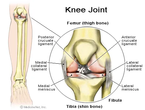
The knee is a joint which has three parts. The thigh bone (femur) meets the large shin bone (tibia) forming the main knee joint. This joint has an inner (medial) and an outer (lateral) compartment. The kneecap (patella) joins the femur to form a third joint, called the patellofemoral joint.
The knee joint is surrounded by a joint capsule with ligaments strapping the inside and outside of the joint (collateral ligaments) as well as crossing within the joint (cruciate ligaments). These ligaments provide stability and strength to the knee joint.
The meniscus is a thickened cartilage pad between the two joints formed by the femur and tibia. The meniscus acts as a smooth surface for the joint to move on. The knee joint is surrounded by fluid-filled sacs called bursae, which serve as gliding surfaces that reduce friction of the tendons. There is a large tendon (patellar tendon) which envelopes the knee cap and attaches to the front of the tibia bone. There are large blood vessels passing through the area behind the knee (referred to as the popliteal space). The large muscles of the thigh move the knee. In the front of the thigh, the quadriceps muscles extend, or straighten, the knee joint by pulling on the patellar tendon. In the back of the thigh, the hamstring muscles flex, or bend, the knee. The knee also rotates slightly under guidance of specific muscles of the thigh.
Knee Joint
The knee functions to allow movement of the leg and is critical to normal walking. The knee flexes normally to a maximum of 135 degrees and extends to 0 degrees. The bursae, or fluid-filled sacs, serve as gliding surfaces for the tendons to reduce the force of friction as these tendons move. The knee is a weight-bearing joint. Each meniscus serves to evenly load the surface during weight-bearing and also aids in disbursing joint fluid for joint lubrication.

What injuries can cause knee pain?

Injury can affect any of the ligaments, bursae, or tendons surrounding the knee joint. Injury can also affect the ligaments, cartilage, menisci (plural for meniscus), and bones forming the joint. The complexity of the design of the knee joint and the fact that it is an active weight-bearing joint are factors in making the knee one of the most commonly injured joints.

Ligament Injury of the Knee

Trauma can cause injury to the ligaments on the inner portion of the knee (medial collateral ligament), the outer portion of the knee (lateral collateral ligament), or within the knee (cruciate ligaments). Injuries to these areas are noticed as immediate pain, but are sometimes difficult to localize. Usually, a collateral ligament injury is felt on the inner or outer portions of the knee. A collateral ligament injury is often associated with local tenderness over the area of the ligament involved. A cruciate ligament injury is felt deep within the knee. It is sometimes noticed with a "popping" sensation with the initial trauma. A ligament injury to the knee is usually painful at rest and may be swollen and warm. The pain is usually worsened by bending the knee, putting weight on the knee, or walking. The severity of the injury can vary from mild (minor stretching or tearing of the ligament fibers, such as a low grade sprain) to severe (complete tear of the ligament fibers). Patients can have more than one area injured in a single traumatic event.


POSTED BY ATTORNEY RENE G. GARCIA:
- Some of our clients have suffered major knee and leg injuries due to a serious accident. The Garcia Law Firm, P.C. was able to successfully handle these types of cases. For a free consultation please call us at 1-866- SCAFFOLD or 212-725-1313.

Link: http://www.webmd.com/osteoarthritis/osteoarthritis-knee-injuries

Knee Problems and Injuries


    Most people have had a minor knee problem at one time or another. Most of the time our body movements do not cause problems, but it's not surprising that symptoms develop from everyday wear and tear, overuse, or injury. Knee problems and injuries most often occur during sports or recreational activities, work-related tasks, or home projects.
The knee is the largest joint in the body. The upper and lower bones of the knee are separated by two discs (menisci). The upper leg bone (femur) and the lower leg bones (tibia and fibula) are connected by ligaments, tendons, and muscles. The surface of the bones inside the knee joint is covered by articular cartilage, which absorbs shock and provides a smooth, gliding surface for joint movement. See a picture of the structures of the knee camera.
Although a knee problem is often caused by an injury to one or more of these structures, it may have another cause. Some people are more likely to develop knee problems than others. Many jobs, sports and recreation activities, getting older, or having a disease such as osteoporosis or arthritis increase your chances of having problems with your knees.

Sudden (acute) injuries

Injuries are the most common cause of knee problems. Sudden (acute) injuries may be caused by a direct blow to the knee or from abnormal twisting, bending the knee, or falling on the knee. Pain, bruising, or swelling may be severe and develop within minutes of the injury. Nerves or blood vessels may be pinched or damaged during the injury. The knee or lower leg may feel numb, weak, or cold; tingle; or look pale or blue. Acute injuries include:
  • Sprains, strains, or other injuries to the ligaments and tendons that connect and support the kneecap.
  • A tear in the rubbery cushions of the knee joint (meniscus).
  • Ligament tears, such as the anterior cruciate ligament (ACL). The medial collateral ligament (MCL) is the most commonly injured ligament of the knee.
  • Breaks (fracture) of the kneecap, lower portion of the femur, or upper part of the tibia or fibula. Knee fractures are most commonly caused by abnormal force, such as a falling on the knee, a severe twisting motion, severe force that bends the knee, or when the knee forcefully hits an object.
  • Kneecap dislocation. This type of dislocation occurs more frequently in 13- to 18-year-old girls. Pieces of bone or tissue (loose bodies) from a fracture or dislocation may get caught in the joint and interfere with movement.
  • Knee joint dislocation. This is a rare injury that requires great force. It is a serious injury and requires immediate medical care.

Overuse injuries

Overuse injuries occur with repetitive activities or repeated or prolonged pressure on the knee. Activities such as stair climbing, bicycle riding, jogging, or jumping stress joints and other tissues and can lead to irritation and inflammation. Overuse injuries include:
  • Inflammation of the small sacs of fluid that cushion and lubricate the knee (bursitis).
  • Inflammation of the tendons (tendinitis) or small tears in the tendons (tendinosis).
  • Thickening or folding of the knee ligaments (plica syndrome).
  • Pain in the front of the knee from overuse, injury, excess weight, or problems in the kneecap (patellofemoral pain syndrome).
  • Irritation and inflammation of the band of fibrous tissue that runs down the outside of the thigh (iliotibial band syndrome).
POSTED BY ATTORNEY RENE G. GARCIA:
- Some of our clients have suffered major knee and leg injuries due to a serious accident. The Garcia Law Firm, P.C. was able to successfully handle these types of cases. For a free consultation please call us at 1-866- SCAFFOLD or 212-725-1313.

Link: http://www.webmd.com/pain-management/knee-pain/knee-problems-and-injuries-topic-overview

Carpal Tunnel Syndrome

Source: http://emedicine.medscape.com/article/1243192-overview

Carpal tunnel syndrome (CTS) is the most commonly diagnosed and treated entrapment neuropathy. The syndrome is characterized by pain, paresthesia, and weakness in the median nerve distribution of the hand. Surgical and nonsurgical treatments exist that can produce excellent outcomes for patients.
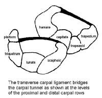
An image depicting the carpal canal can be seen below.
Cross sections of the carpal canal at the levels oCross sections of the carpal canal at the levels of the proximal and distal carpal rows are depicted. The transverse carpal ligament bridges the carpal tunnel and is under tension.

American Academy of Orthopaedic Surgeons guidelines

The AAOS has developed the following clinical guidelines for the treatment of carpal tunnel syndrome (level of evidence provided in parentheses):[1]
Recommendation 1
A course of nonoperative treatment is an option in patients diagnosed with carpal tunnel syndrome (CTS). Early surgery is an option when there is clinical evidence of median nerve denervation or the patient elects to proceed directly to surgical treatment. (Grade C, level V)
Recommendation 2
We suggest another nonoperative treatment or surgery when the current treatment fails to resolve the symptoms within 2-7 weeks. (Grade B, level I and II)
Recommendation 3
We do not have sufficient evidence to provide specific treatment recommendations for carpal tunnel syndrome when found in association with the following conditions: diabetes mellitus, coexistent cervical radiculopathy, hypothyroidism, polyneuropathy, pregnancy, rheumatoid arthritis, and carpal tunnel syndrome in the workplace. (Inconclusive, No evidence found)
Recommendation 4a
Local steroid injection or splinting is suggested when treating patients with carpal tunnel syndrome, before considering surgery. (Grade B, level I and II)
Recommendation 4b
Oral steroids or ultrasound are options when treating patients with carpal tunnel syndrome. (Grade C, level II)
Recommendation 4c
We recommend carpal tunnel release as treatment for carpal tunnel syndrome. (Grade A, level I)
Recommendation 4d
Heat therapy is not among the options that should be used to treat patients with carpal tunnel syndrome. (Grade C, level II)
Recommendation 4e
The following treatments carry no recommendation for or against their use: activity modifications, acupuncture, cognitive behavioral therapy, cold laser, diuretics, exercise, electric stimulation, fitness, Graston instrument, iontophoresis, laser, stretching, massage therapy, magnet therapy, manipulation, medications (including anticonvulsants, antidepressants, and nonsteroidal anti-inflammatory drugs [NSAIDs]), nutritional supplements, phonophoresis, smoking cessation, systemic steroid injection, therapeutic touch, vitamin B6 (pyridoxine), weight reduction, yoga. (Inconclusive, level II and V)
Recommendation 5
We recommend surgical treatment of carpal tunnel syndrome by complete division of the flexor retinaculum regardless of the specific surgical technique. (Grade A, level I and II)
Recommendation 6
We suggest that surgeons do not routinely use the following procedures when performing carpal tunnel release: skin nerve preservation (Grade B, level I); epineurotomy (Grade C, level II)
The following procedures carry no recommendation for or against use: flexor retinaculum lengthening, internal neurolysis, tenosynovectomy, ulnar bursa preservation. (Inconclusive, level II and V)
Recommendation 7
The physician has the option of prescribing preoperative antibiotics for carpal tunnel surgery. (Grade C, level III)
Recommendation 8
We suggest that the wrist not be immobilized postoperatively after routine carpal tunnel surgery. (Grade B, level II)
We make no recommendation for or against the use of postoperative rehabilitation. (Inconclusive, level II)
Recommendation 9
We suggest physicians use one or more of the following instruments when assessing patients’ responses to CTS treatment for research:
  • Boston Carpal Tunnel Questionnaire (disease-specific)
  • DASH—Disabilities of the Arm, Shoulder, and Hand (region-specific; upper limb)
  • MHQ—Michigan Hand Outcomes Questionnaire (region-specific; hand/wrist)
  • Patient Evaluation Measure (region-specific; hand)
  • SF-12 or SF-36 Short Form Health Survey (generic; physical health component for global health impact) (Grade B, level I, II, and III)

Recent studies

Jarvik et al compared surgical (57 patients) versus multi-modality, nonsurgical treatment (hand therapy and ultrasound; 59 patients) for carpal tunnel syndrome without denervation. Analyses showed a significant 12-month adjusted advantage for surgery in function and symptoms; there were no clinically important adverse events and no surgical complications. According to the authors, symptoms in both groups improved, but surgical treatment led to better outcome than nonsurgical treatment.[2]
In a Mayo Clinic study by Gelfman et al, temporal trends in CTS were assessed for incidence, surgical treatment, and lost time at work. Using Olsmsted County residents as the study population, 10,069 residents were found to have been diagnosed with CTS from 1981-2005 (491 per 100,000 person years for women; 258 per 100,000 person years for men; 376 per 100,000 person-years combined). Adjusted annual rates increased from 258 per 100,000 in 1981-1985 to 424 in 2000-2005. The average annual incidence of carpal tunnel release surgery was 109 per 100,000, and that for work-related CTS was 11 per 100,000. According to the authors, the increase seen in this population corresponds to a national epidemic of CTS cases resulting in lost work days that began in the mid-1980s and lasted through the mid-1990s, but the cause for the increase is not yet clear.[3]
Pomerance et al compared direct costs and results for patients with electrodiagnostically proven CTS treated with surgery versus nonsurgical care. In the study, 120 patients were divided into 2 groups: group 1 received nonsurgical therapy, and group 2 received surgical treatment. Follow-up averaged 13 ± 5 months for group 1 and 12 ± 2 months for group 2, with 32 patients in group 1 electing to have surgery during the follow-up period. Cost of care averaged $3,335 ± $2,097 in the nonsurgery group and $3,068 ± $983 in the surgery group. The authors concluded that surgery should be considered as the initial form of treatment when patients are diagnosed with CTS confirmed by nerve conduction studies because surgery provides symptom resolution with a favorable cost analysis.[4]
Wolf et al studied the diagnosis of CTS in the United States military population from 1998-2006 and found the incidence to be 3.98 per 1,000 person-years, which compared with incidences of 1.5 to 3.5 per 1,000 person-years in other regional or working-group populations studied. In the military study, females had a significantly higher incidence of CTS than males, with an adjusted incidence rate ratio of 3.29. CTS incidence was found to increase with age, with the age group 40 years or older having a significantly higher incidence. Additionally, military rank was found to be an independent risk factor for CTS, with rates higher in senior officer and enlisted groups, suggesting that occupational requirements have an effect on CTS within the military.[5]
For excellent patient education resources, visit eMedicine's Hand, Wrist, Elbow, and Shoulder Center and Arthritis Center.

*Some of our clients have suffered impaired hand and forearm trauma due to a serious accident. The Garcia Law Firm, P.C. was able to successfully handle these types of cases. For a free consultation please call us at 1-866- SCAFFOLD or 212-725-1313. edicine's patient education article Carpal Tunnel Syndrome.

Carpal Ligament Instability

Source: http://emedicine.medscape.com/article/1241610-overview

The human wrist joint is a complex arrangement of small bones and ligaments that form a mobile yet stable link from the powerful forearm to the hand. The normally functioning carpus can position the hand precisely relative to the forearm and provides remarkably stable transmission of forces. Motion and stability of the carpus provide the critical foundation for maximum hand function from precise fine motor control to power grip activities.
When the normal mechanics of the wrist are disrupted, the instability of the carpal bones results in weakness, stiffness, chronic pain, and often arthritis if not treated appropriately. Although the early clinical and radiographic findings may be subtle, an understanding of wrist kinematics and instability patterns can facilitate early diagnosis and management. Unfortunately, selecting the optimal treatment remains a difficult judgment in most cases.
Linscheid et al described traumatic carpal instability in 1972.[1] Since the early reports, anatomic and biomechanical studies have provided a foundation for understanding carpal motion, stresses, and pathologic instability. Building on these studies, various models have been suggested to explain the remarkable strength and mobility of this complex joint and the predictable patterns of failure.
This article presents the current understanding of pathologic carpal instability, the common classification patterns, and early treatment options that may avoid protracted dysfunction. Appropriate hand therapy is essential to maximize recovery but requires an appreciation of the limitations of carpal instability dysfunction and the goals of various treatment options.
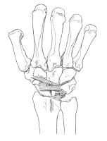
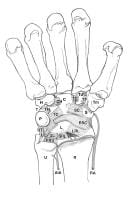
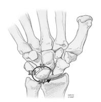
Images of the carpal ligament and instability are included below.
(Click image to enlarge.) Dorsal carpal ligaments.(Click image to enlarge.) Dorsal carpal ligaments. Copyright Mayo Clinic, used with permission of Mayo Foundation. (Click image to enlarge.) Volar carpal ligaments. (Click image to enlarge.) Volar carpal ligaments. Copyright Mayo Clinic, used with permission of the Mayo Foundation. Mayfield perilunate instability pattern. CopyrightMayfield perilunate instability pattern. Copyright Mayo Clinic, used with permission of the Mayo Foundation.

 -- Some of our clients have suffered impaired hand and forearm trauma due to a serious accident. The Garcia Law Firm, P.C. was able to successfully handle these types of cases. For a free consultation please call us at 1-866- SCAFFOLD or 212-725-1313.

Hand SurgeryImprove Strength, Function and Flexibility

       Source: http://www.plasticsurgery.org/Reconstructive-Procedures/Hand-Surgery.html

Hand surgery can treat diseases that cause pain and impair the strength, function and flexibility of your wrist and fingers.

    Types of hand surgery

     If your hand is impaired in any way, surgery may improve your condition. This type of very specialized surgery can treat diseases that cause pain and impair the strength, function and flexibility of your wrist and fingers. Surgery seeks to restore to near normal the function of fingers and hands injured by trauma or to correct abnormalities that were present at birth. Specifically, hand surgery can treat:
    Carpal tunnel syndrome – a condition caused by pressure to the median nerve within the wrist, or carpal tunnel. You might feel pain, a tingling sensation, numbness of the fingers, weakness or aching. Carpal tunnel syndrome is associated with multiple conditions including: repetitive motion or overuse, fluid retention during pregnancy, injury to the nerve in the carpal tunnel or rheumatoid arthritis.
    Rheumatoid arthritis – a disabling disease that can cause severe inflammation in any joint of the body. In the hand, it can deform fingers and impair movement.
    Dupuytren’s contracture – a disabling hand disorder in which thick, scar-like tissue bands form within the palm and may extend into the fingers. It can cause restricted movement, bending the fingers into an abnormal position.

    Is it right for me?

    Hand surgery is a highly individualized procedure which can be performed on people of any age and is a good option for you if:
    • You do not have additional medical conditions or other illnesses that may impair healing
    • You are a non-smoker
    • You have a positive outlook and realistic goals for your hand surgery
    • You are committed to following your plastic surgeon’s prescribed course of treatment
    • In some conditions, hand surgery is necessary to treat wounds and to help painful conditions.

     -- Some of our clients have suffered impaired hand and forearm trauma due to a serious accident. The Garcia Law Firm, P.C. was able to successfully handle these types of cases. For a free consultation please call us at 1-866- SCAFFOLD or 212-725-1313.

    Spinal Cord Stimulators - the 'pacemaker' for chronic pain

                                         Source:http://www.gizmag.com/eon-mini-spinal-cord-stimulator/12486/

    Spinal Cord Stimulators - the 'pacemaker' for chronic pain

    By
    August 12, 2009


    The Eon Mini Spinal Cord Stimulator
    The Eon Mini Spinal Cord Stimulator
    Image Gallery (4 images)
    Free Knee Pain Relief Kit - Osteoarthritis-Relief.com
    Get Free Info On Osteoarthritis Knee Pain Treatment Today.
    Infuse Lawsuit - www.YourLawyer.com
    Severe Bone Pain & Swelling Due To Infuse Bone Growth? Get Legal Help!
    Sciatica - Pain Doctor - NYSpineMedicine.com
    End back pain without surgery Dr. Douglas Schottenstein, MD
    Find a Doctor Near You - TurnToHelp.com
    Opioid Dependence Treatment in the Privacy of a Doctor's Office
    Ads by Google
    While nobody's exactly sure how it works, it's been clinically proven over the past 30-40 years that low levels of electrical energy, delivered straight to the nerve fibers in the spinal cord, can disrupt the signals that certain chronic pain conditions send to the brain, and replace them with a much more pleasant tingling sensation. Now, St Jude Medical has received FDA and CE mark approvals for the world's smallest and longest-lasting rechargeable neurostimulator. The Eon Mini is the size of a silver dollar, about 1cm thick (1/2 inch), and weighs only 29g (1oz). It sits under the skin of the buttock or abdomen, and its rechargeable battery should last nearly 10 years. It can be programmed by remote control to treat as many as eight different chronic pain areas and, in doing so, it can get many patients with chronic pain off morphine and back into a semblance of normal life.
    Chronic pain is a broad condition that simply refers to pain that continues to be present long after an injury is expected to be healed. For many sufferers, it's a crippling and ever-present fact of life that affects work, relationships, sleep and many other areas. Often, the side effects of pain relief strategies can be as crippling as the pain itself.
    The first use of small electrical currents as a human analgesic was reported in 1971 - a Japanese team found that by sending small electrical currents through wires running along the epidural space - the outermost part of the spinal canal - they could disrupt the crippling pain signals being sent back to the brain, and replace them with a much more pleasant buzzing sensation.
    Spinal Cord Stimulation (SCS) is not a cure for chronic pain conditions - it's just a pain relief strategy. But it can be extremely effective, reducing the pain by 50 percent or more without the side effects associated with opioid analgesics like morphine. It's viewed as somewhat of a last resort for cases in which patients are unable to achieve a satisfactory quality of life using other pain relief methods.
    The Eon Mini represents a step forward in the delivery of SCS pain relief. Its small size allows the main unit to be implanted more deeply and less obtrusively under the skin than larger alternatives, and the 10-year life of its rechargeable battery means that patients need replacement operations far less frequently.
    The patient first takes a short trial to see if the system is likely to deliver effective pain relief. This involves the implantation of the electrodes, but not the SCS device. If the doctor deems the results satisfactory and the patient suitable, the Eon Mini is buried under the skin.
    The doctor is able to treat up to eight pain areas through 16 different header contacts, allowing the treatment of complex pain patterns throughout the lower body. It's programmed on the operating table with the patient awake and reporting back to the doctor on which areas of pain need the most attention. It's usually an outpatient treatment, and the results are immediate and last as long as the battery.
    It's not precisely known how the SCS technique confuses the body's pain receptors - it could be one of a handful of different ways. But it's effective in many cases, and shows very few side effects. More information at www.poweroveryourpain.com.

    -- Some of our clients have suffered chronic back pain due to a serious accident. The Garcia Law Firm, P.C. was able to successfully handle these types of cases. For a free consultation please call us at 1-866- SCAFFOLD or 212-725-1313.